Radiology
Complete Radiology Services under one roof
In the world of diagnostics, our Radiology Center stands as a beacon of excellence, offering a comprehensive range of imaging services—all conveniently housed under one roof. From X-rays to CT scans, sonography to mammography, our state-of-the-art facility is dedicated to providing you with a seamless and thorough radiological experience. Embrace the convenience of having all your imaging needs met with precision and care, ensuring a comprehensive approach to your healthcare journey. Welcome to a Radiology Center where excellence meets convenience, bringing you complete radiological services under one roof.
Evaluates cardiac function during exercise, providing insights into heart health
and potential cardiovascular issues.
Records the heart’s electrical activity, aiding in the diagnosis of various cardiac conditions.
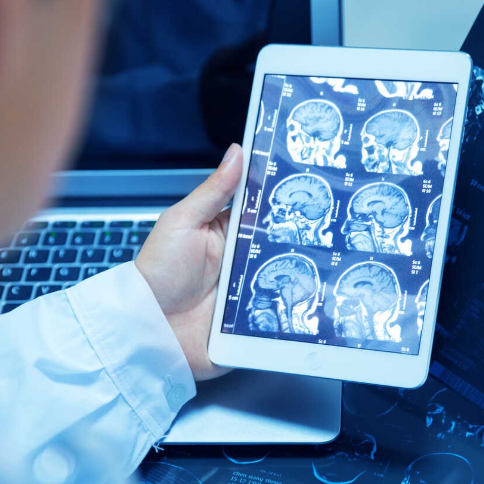
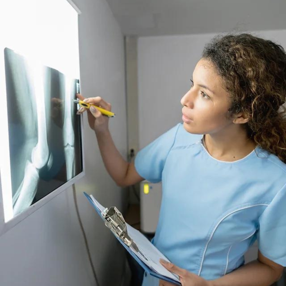
Utilizes electromagnetic radiation to produce images of internal structures, assisting in the diagnosis of bone and certain soft tissue conditions.
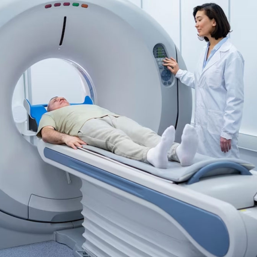
Generates detailed cross-sectional images of the body, enabling comprehensive examination of internal structures for diagnostic purposes.
Uses sound waves to create images of organs and tissues, offering a non-invasive method for assessing various medical conditions.
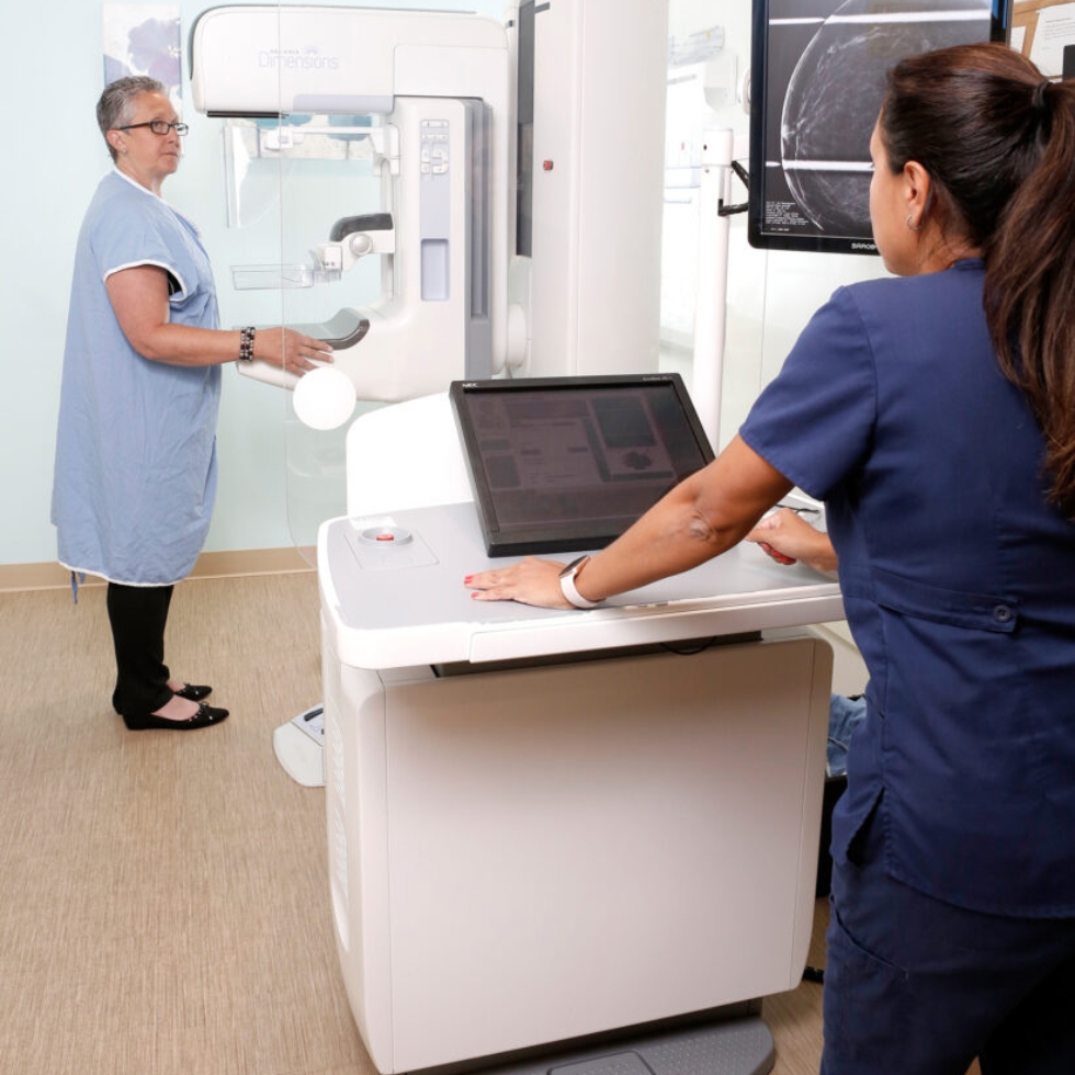
Specialized X-ray imaging for breast examination, essential in detecting and diagnosing breast cancer at early, treatable
Digital X-Ray
Digital X-Ray technology has transformed the landscape of diagnostic imaging, providing numerous benefits in efficiency and diagnostic accuracy. Unlike traditional X-rays, Digital X-Ray uses electronic sensors to capture and store images digitally. This not only reduces radiation exposure for patients but also allows for quick image retrieval and manipulation.
One significant advantage is the speed of image acquisition. Digital X-Ray produces images in seconds, expediting the diagnostic process and enabling swift medical intervention. The digital format facilitates easy storage, retrieval, and sharing of images, enhancing collaboration among healthcare professionals.
Moreover, Digital X-Ray images can be manipulated to improve visibility of specific areas, aiding radiologists in making more accurate diagnoses. The technology’s compatibility with Picture Archiving and Communication Systems (PACS) streamlines the storage and retrieval of patient records, contributing to a more organized and accessible healthcare environment.
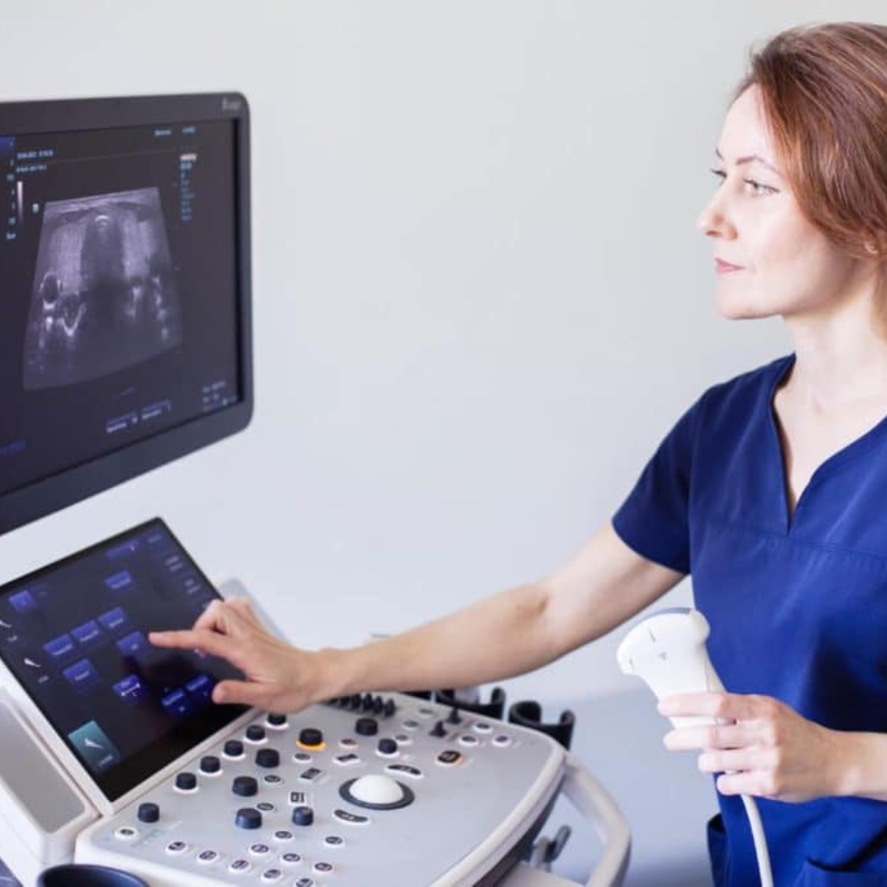
Sonography
Sonography, also known as ultrasound imaging, has become a cornerstone in diagnostic medicine due to its non-invasive and versatile nature. This medical imaging technique utilizes high-frequency sound waves to create detailed images of the internal structures of the body.
One of the primary advantages of sonography is its safety, as it doesn’t involve ionizing radiation. This makes it particularly useful in monitoring fetal development during pregnancy. Obstetric ultrasound provides crucial insights into the health and growth of the fetus, aiding healthcare professionals in ensuring a safe pregnancy.
Beyond obstetrics, sonography plays a vital role in various medical fields. Abdominal sonography helps visualize organs such as the liver, kidneys, and gallbladder, assisting in the diagnosis of conditions like kidney stones and liver diseases. Additionally, cardiac ultrasound, or echocardiography, provides detailed images of the heart’s structure and function, aiding in the assessment of cardiovascular health.
Mamography
Mammography, a specialized X-ray imaging technique for the breast, is a pivotal tool in the early detection and screening of breast cancer. This non-invasive procedure plays a crucial role in women’s health by capturing detailed images of breast tissue to identify abnormalities that may indicate the presence of cancerous or precancerous cells.
One of the primary advantages of mammography is its ability to detect breast abnormalities at an early stage, often before they can be felt during a physical examination. Early detection significantly improves the chances of successful treatment and a positive prognosis for breast cancer patients.
Digital mammography, an advancement in this field, allows for the electronic capture and storage of images, offering enhanced clarity and diagnostic accuracy. Computer-aided detection (CAD) systems further assist radiologists by highlighting potential areas of concern, improving the overall efficiency of the screening process.
BMD
Bone Mineral Density (BMD) testing is a critical diagnostic tool used in assessing bone health and detecting conditions such as osteoporosis. This non-invasive procedure measures the amount of mineral content in bones, providing valuable insights into bone strength and density.
BMD testing is commonly performed using Dual-Energy X-ray Absorptiometry (DEXA) scans, which are highly accurate and widely considered the gold standard in bone density assessment. During the test, low-dose X-rays are directed at specific bones, typically the spine, hip, or forearm. The amount of X-ray energy absorbed by the bones is then measured, indicating their mineral density.
The results of BMD testing help healthcare professionals evaluate a patient’s risk of fractures and assess overall bone health. Individuals with lower BMD, especially postmenopausal women and older adults, may be at a higher risk of fractures and osteoporosis. Early detection through BMD testing allows for timely intervention and preventive measures, such as lifestyle changes, medications, and dietary adjustments.
Colour Doppler
Color Doppler imaging is a specialized ultrasound technique that provides valuable insights into blood flow within the body’s vessels. By combining traditional ultrasound with color mapping, this diagnostic tool enables healthcare professionals to visualize and assess blood circulation in real-time.
Primarily used in vascular studies, Color Doppler helps identify abnormalities in blood flow, such as blockages, narrowing of vessels, or abnormal blood clots. The color-coded images generated by this technology allow clinicians to distinguish between blood moving toward and away from the transducer, providing a dynamic representation of blood flow patterns.
One of the significant applications of Color Doppler is in cardiovascular assessments, aiding in the evaluation of conditions such as arterial and venous diseases. Cardiologists use this imaging modality to examine the blood vessels around the heart and detect potential issues like stenosis or aneurysms.

Helpful Links
Get in touch with us!
Contact: 7887830000/7410047378
Email:
info@suyoglifecare.com
Address:
Office no.10 Suyog Center, Gultekdi, Market Yard Road, Pune-411037.